Handheld 3D Ophthalmic Ultrasound Tomography
Designing and building a device to standardize the acquisition and generation of 3D ophthalmic tomography models.
EXPLORE THE PROJECT
Develop a handheld device that standardizes the acquisition and generation of 3D ophthalmic tomography models.
Models can be used to delineate the vitreous, retina and optic nerve and could be used for quantitative measurements of gross ophthalmic anatomy.
Create a 3D reconstructive technique so that patients and physicians can better spatially visualize ocular pathology and possibly quantify depth and volume changes.
PROJECT GOALS
This is a handheld, battery operated, ultrasound rotation point of care (POC) device that standardizes the way ophthalmic ultrasound images are taken. This device mechanically rotates a handheld Butterfly iQ ultrasound probe across the human probe to acquire images of the globe. Videos and images of the globe are converted into image slices that are then stitched together in a computer to render a 3D image of the anatomical structures of the eye, which can be used for evaluation and diagnosis. This device can be used at the bedside, in the OR, or in any low-resource setting.
HOW IT WORKS
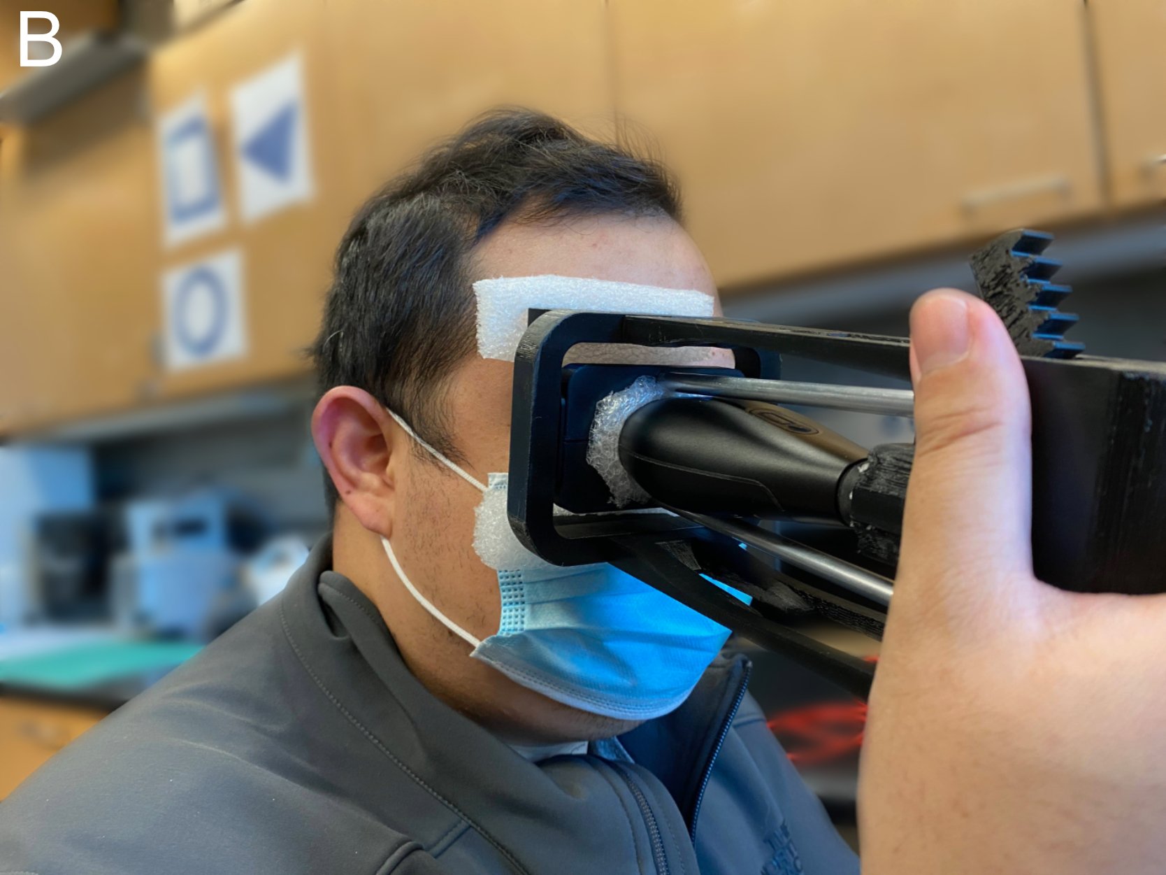
Demonstration of Use
The device is held over a human subject’s eye during ultrasound imaging.
Clinical Workflow
These are the steps of how the device works, rotating a US probe across a human eye and then converting captured 2D image data into a 3D model.
I designed and modeled the parts in Fusion 360, printed the parts on a Prusa MKS3+, programmed the arduino to move a servo motor, and assembled a fully functional prototype.
HOW ITS BUILT
In collaboration with Professor Tang’s students we wrote a custom python-matlab processing program. Our program takes the images acquired from the ultrasound, enhances the images, interpolates pixel data between image slices, and then constructs a custom .nii 3D model file. The 3D model can be opened in an open source software used by the medical imaging community called, iTK- SNAP. Alternatively the 3D file can be opened in 3D Slicer.
RESULTS
Ultrasound Images
3D Model
This model of my eye was created using images captured by the device.
This project could still use some work, and I plan to continue to tweak and improve the design. My goals are to:
FUTURE GOALS
1. Redesign handheld form factor
Incorporate power switch and other programmable switches
Encapsulate entire ultrasound probe inside a case to hide/protect mechanism
Improve ease of recharging butterfly ultrasound probe by either improving removal of probe from device or modifying device so the probe does not have to be removed
Streamline data acquisition with hand held ultrasound acquisition device with 3D tomography construction.
2. Streamline data acquisition with 3D tomography construction.
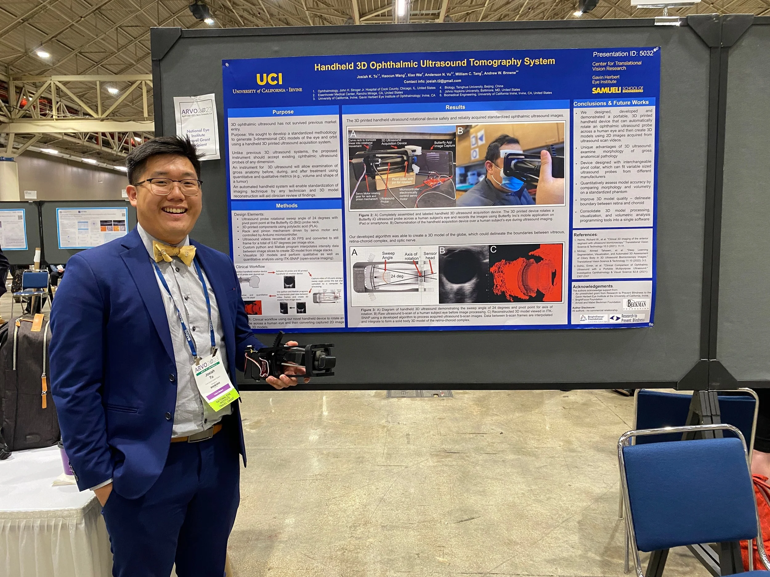
PRESENTING THE WORK
Our device is currently under patent review and can be viewed here.
I am also extremely grateful to the National Eye Institute (NEI) for awarding me a grant to present this work at the 2023 American Research for Vision and Ophthalmology (ARVO) conference in New Orleans.